Ocular Anatomy and Refractive Error
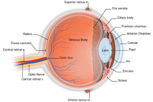
The eye is a truly amazing organ. Just the size of a ping-pong ball, this complex little globe converts the light around us into vibrant images. While the anatomy of the eye is somewhat complex, its overall function is much like that of a camera. This section will explain how the normal eye works and how refractive errors such as nearsightedness and farsightedness develop.

Next, light passes through the lens of the eye, which sits just behind the iris. The lens further focuses the light, which continues on through the clear vitreous which fills the majority of the eye. Light finally arrives at the retina, a tissue paper thin membrane which lines the inside of the eyeball. The retina is the eye’s film, sensing the light we see and converting it into signals which are then sent on to the brain via the optic nerve. The brain, like a computer, further processes information from the retina, creating the images we see.
In the normal eye, distant light rays are precisely focused by the cornea and lens onto the retina, leading to excellent visual acuity, generally 20/20 or better. (This numerical description of visual acuity means that the person being tested can see at 20 feet what a normal sighted person can see at 20 feet. The limit of normal human visual acuity has been determined by study of the eye.) If there is an imbalance between the power of the refractive structures of the eye (cornea and lens) and the eye’s length, light will not be properly focused on the retina. Depending on the type of imbalance, either nearsightedness, known as myopia, or farsightedness, known as hyperopia, occurs. These refractive errors are measured in units known as diopters .
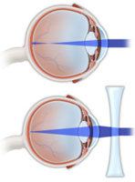
Nearsightedness (myopia) develops if the cornea/lens power is too strong, or the eye is too long. In this case, a person can see well at near, but distant objects are out of focus. Corrective lenses refocus the light onto the retina in order to improve distance visual acuity (see diagram at left). Refractive surgery either reshapes the cornea or uses an implanted lens to refocus light properly onto the retina to achieve good vision.
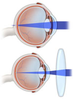
HyperopiaFarsightedness (hyperopia) occurs if the cornea/lens power is too weak, or if the eye is too short. This leads to poor near vision and better distance vision, though in some cases vision is not clear at any distance without correction (see diagram at right). Again, corrective lenses or refractive surgery can refocus light to achieve good acuity.
Astigmatism develops when the curvature of the cornea or lens is irregular, leading to different refractive powers depending upon the direction of the incoming light. This condition also leads to blurring of vision and can be corrected with lenses or surgery.
Presbyopia is the age-related loss of the ability to change focus from far to near. Most people notice its onset between 40 and 45 years of age. Before the onset of presbyopia, the normal human lens can change shape as muscles inside the eye adjust, allowing continually adaptive focusing of the eye.
As presbyopia develops and progresses, the flexibility of the lens and muscles diminishes, and reading glasses or bifocals become necessary for near viewing. Presently, one FDA-approved procedure, conductive keratoplasty (CK) is available specifically for the treatment of presbyopia. This will be discussed later.
Free Lasik Screening
Call 520-355-7501 or Enter Your Information Below!
Please do not submit any Protected Health Information (PHI).
More Categories


