Fishkind, Bakewell, Maltzman, Hunter, & Associates is now Oracle Eye Physicians & Surgeons!

Providing the same excellent eye care because It Matters Who You See!

Skip to main content
Mon - Fri: 8:00 am - 5:00 pm, Sat & Sun: Closed
Give Us A Call Today!
(520) 355-7501
Give Us A Call Today!
(520) 293-6740

Author: eyestucson

Glaucoma : Angle Closure and Narrow Angles

Narrow Angles and Angle Closure Glaucoma
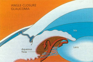
Closed angle glaucoma is relatively uncommon compared with the typical open angle form. In open angle glaucoma the entrance into the trabecular meshwork is unobstructed, though an abnormality deep within the meshwork leads to elevated intraocular pressure. In closed angle glaucoma, the entrance to the trabecular meshwork becomes blocked by the iris, glaucoma4 which assumes an abnormally forward position. Aqueous humor cannot gain access to the trabecular meshwork and drainage canals, causing the intraocular pressure to increase.

People with closed angle glaucoma often have a shorter than average eye in which all the internal structures, including the cornea and the iris, are closer together than normal. Many of these individuals are hyperopic, or farsighted. Others have a condition called plateau iris, in which the iris is attached too far forward in the eye. With age, the eye’s lens thickens, pushing the iris further forward and narrowing the angle. Eventually the angle can become completely blocked by the peripheral iris, preventing the flow of aqueous humor from the eye. This leads to elevated intraocular pressure and optic nerve damage.

Unlike symptom-free open angle glaucoma, this condition, sometimes called “narrow angle glaucoma” or “acute angle closure,” often presents suddenly, with pain, red eye, tearing, and colored haloes around lights. Vision can be lost quickly, and urgent treatment is required. In other cases the closure of the angle is slow and progressive, leading to a condition more like open angle glaucoma, with slow damage and no symptoms.

Risk Factors for Angle Closure Glaucoma

  • Hyperopia. Being farsighted is a major risk factor for narrow angles and angle closure. Hyperopic eyes tend to be short, causing crowding of the angle structures and narrowing the angle.
  • Race. Many Asians and Eskimos have shallow anterior chambers, predisposing them to angle closure.
  • Age. As we age, the lens becomes thicker and larger, increasing the risk of angle closure in predisposed individuals.
  • Sex. Women are 2-3 times more likely than men to suffer angle closure.

Treating Narrow Angles/Angle Closure Glaucoma

Narrow angles and angle closure glaucoma, while often treated with some of the same medications and surgical procedures as open angle glaucoma, have one notable difference. In these conditions, the problem of a narrow angle caused by the iris and cornea being too close together is often exacerbated by a situation known as pupillary block.
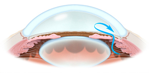
Remember that aqueous humor, the fluid in the front of the eye, is produced behind the iris and must pass through the pupil before gaining access into the trabecular meshwork.

Flow of aqueous humor through a peripheral iridotomy Due to resistance of flow at the pupil , there is actually a slightly higher pressure behind the iris than in front, and this tends to bow the iris forward, contributing to the already narrow angle, and further blocking outflow of fluid from the eye. This papillary block is commonly the cause of acute angle closure in predisposed individuals. Treatment is aimed at alleviating this condition.

A laser is used to create a small opening (or openings) in the periphery of the iris. Known as laser peripheral iridotomy, this procedure creates a lower resistance pathway for aqueous to pass from behind to in front of the iris, preventing papillary block and significantly reducing or eliminating the risk of acute closure of the angle. Iridotomy is effective and safe, with little risk of significant complication. The same procedure is required emergently to treat an actual attack of angle closure, however is much more difficult to perform and may be too late to prevent permanent damage to the eye. Therefore, if your physician finds narrow angles, laser iridotomy may be recommended on a prophylactic basis.

Glaucoma: A General Overview of the Disease

What is Glaucoma?

Glaucoma is a group of eye disorders that can lead to progressive loss of vision, often without any symptoms. Visual loss results from damage to the optic nerve, which transmits signals from the eye to the brain much like an electrical cable. Once nerve fibers are damaged by glaucoma, the information they supply can no longer reach the brain. Peripheral vision is usually lost first. Because central vision is typically preserved until late in the disease and pain is usually absent, most people with glaucoma don’t realize that anything is wrong. Optic nerve damage and vision loss due to glaucoma cannot be reversed, making early diagnosis and initiation of treatment very important.

It is estimated that as much as one to two percent of the US population has open angle glaucoma, and that half of those affected have not been diagnosed. It was once believed that glaucoma is caused by high pressure within the eye, known as intraocular pressure, or IOP. While IOP plays a large role in glaucoma, it is now considered a major risk factor, though not necessarily the specific cause of the disease. The higher the pressure the more likely one is to get glaucoma. However, up to 50 percent of patients with glaucoma have normal intraocular pressures, indicating that other factors must be involved. Genetics, nutrition, blood flow, and toxins may all play a part, though details are currently not well understood. Extensive research is being conducted to identify these factors.

Types of Glaucoma

Glaucoma is usually broken down into two major categories: open angle glaucoma and closed angle glaucoma. To understand the difference, we must first know a bit about how eye pressure develops. The eye is essentially a hollow sphere filled with fluid. In the front section of the eye, this fluid is called the aqueous humor. Aqueous humor is produced by specialized cells behind the iris, the colored part of the eye. Fluid then travels through the pupil, the central opening in the iris, to enter the anterior chamber, which is the space in front of the iris and behind the cornea, the clear window on the front of the eye. The cornea and iris meet peripherally to form the drainage angle of the eye. Within this angle is a structure known as the trabecular meshwork, which is the sieve-like entrance to the drainage system of the eye. The aqueous humor passes through this mesh of tissue, exits the eye through a drainage canal, and is collected by veins to return to the bloodstream.

The physician examining your eye can see into the angle using a special mirrored lens. In open angle glaucoma, there is plenty of space between the iris and the cornea, and the trabecular meshwork is unobstructed.

Open angle glaucoma can be further divided into primary and secondary forms. In primary open angle glaucoma, despite the fact that the entrance to the drain is visibly open, there is resistance to the outflow of aqueous humor, as if the drain is clogged somewhere inside. The exact abnormality is not fully understood but is thought to be due to a defect in the trabecular meshwork. The vast majority of people with glaucoma have this form of the disease.

Secondary open angle glaucoma occurs when another ocular disease or condition causes increased resistance to the outflow of fluid. Two common types of secondary open angle glaucoma, pseudo-exfoliation glaucoma and pigmentary dispersion syndrome, are discussed in greater detail in separate articles.

In closed angle glaucoma, the entrance to the trabecular meshwork becomes blocked by the iris, which is positioned abnormally forward, toward the cornea. People with primary closed angle glaucoma often have a shorter than average eye in which all the internal structures, including the cornea and the iris, are closer together than normal. With age and the development of cataracts, the front of the eye becomes even more crowded, exacerbating this underlying issue.

This situation causes the drainage angle to become narrow and eventually to close, blocking the flow of aqueous humor from the eye. Unlike symptom-free open angle glaucoma, this condition, sometimes called “narrow angle glaucoma” or “angle closure glaucoma,” often presents suddenly, with pain, red eye, tearing, and colored haloes around lights. Vision can be lost quickly, and urgent treatment is required. The goal is to discover the patient with a narrow angle early and to provide treatment, thus preventing an attack of angle closure. As with open angle glaucoma, secondary closed angle glaucoma is caused by other diseases within the eye.

Risk Factors for Glaucoma

A number of risk factors for the development of glaucoma have been identified:

  • Intraocular pressure, as already mentioned, is presently the most significant. Intraocular pressure normally runs between 9 and 21 millimeters of mercury. IOP above 21 is considered high, and with increasing pressure comes increasing risk of glaucoma. However, not all high pressure will lead to glaucoma. Furthermore, as mentioned earlier, glaucoma occurs frequently in the presence of normal levels of IOP. Therefore, intraocular pressure alone cannot be used to diagnose the disease, and must be evaluated as part of the entire clinical picture.
  • Race is another significant factor. African Americans are five to six times more likely than Caucasians to be affected by the disease, which tends to be more severe and progress more quickly in this population. Latinos are also at greater risk than Caucasians. The reasons for these racial differences are not well understood, but are most likely genetic.
  • Family history of glaucoma, particularly in a first degree relative, i.e. parent or sibling, increases one’s risk. The genetics of glaucoma, however, are usually not simple, and there is no way to predict which family members will be affected. At the time that this article was last updated, there have been at least 20 genes identified as causing or contributing to a form of glaucoma, with new genes being discovered fairly frequently. Within the next 5 to 10 years, genetic testing may become routinely available to aid in the diagnosis and prognosis of glaucoma.
  • Age: Glaucoma is uncommon under 40, but risk increases with each decade of life.
  • High blood pressure has been associated with increased intraocular pressure in some studies, but it remains unclear exactly how significant this association is clinically.
  • Low blood pressure may be a risk factor for glaucoma progression, particularly in patients with low tension glaucoma, a form of open angle glaucoma in which the pressure is in the normal range, often quite low. The key seems to be the diastolic perfusion pressure- the pressure experienced by the tiny blood vessels of the optic nerve at the end of each heartbeat, measured as the diastolic blood pressure (the second number in your blood pressure measurement) minus the intraocular pressure. Diastolic perfusion pressures below 50 appear to be associated with increased risk of optic nerve damage and glaucoma. This is currently an area of very active study.
  • Sleep-associated breathing disorders, such as sleep apnea, may also be a risk factor, particularly for low tension glaucoma, again due to abnormal blood flow to the optic nerves.

Other possible risk factors include diabetes and myopia (nearsightedness), however studies have provided inconsistent results as to the degree of association with these factors.

Diagnosing Glaucoma

Your physician will perform a complete medical history and eye examination in order to determine your risk factors and to search for signs of disease. The entire eye will be carefully evaluated, with special attention paid to the optic nerve, the structure damaged by glaucoma. The optic nerve is viewed through your dilated pupil using a special lens and an ophthalmic microscope. Gonioscopy may also be performed, in which a special mirrored lens in placed on your eye in order to directly examine the drainage angle and trabecular meshwork. The thickness of your cornea may also be measured, as an abnormally thick or thin cornea can affect the accuracy of intraocular pressure measurement. After your exam, if glaucoma is suspected, additional tests may be recommended, such as:

  • Visual field testing is conducted to evaluate the function of the optic nerve. As glaucoma damage progresses vision is lost, typically beginning in the periphery and moving toward the center. Computerized visual field devices flash a series of lights in your central and peripheral vision in order to map out any areas of visual loss. Again, by repeating this test over a period of time, stability or progression of disease can be ascertained and treatment adjusted as necessary. Completion of this test can take anywhere between 15 and 30 minutes, depending upon the exact type of examination your doctor orders. Your eyes will not be dilated for a visual field.
  • Ocular Coherence Tomography, or OCT, uses a laser to create a three-dimensional image of the optic nerve and surrounding retina, which is then analyzed by a computer. This allows your physician to better evaluate the structure of the fibers of the optic nerve, which are altered by glaucoma. Changes in the structure of the optic nerve occur early in glaucoma, often preceding any visual changes, and identification of these changes may be the best way to diagnose disease. By repeating this exam over time, the success of glaucoma treatment can be monitored. Performance of this test usually does not require dilation and takes about 10 minutes.

Unfortunately, the diagnosis is glaucoma is not always straightforward, even after all testing is performed. There is significant variation in the appearance of normal optic nerves, with many healthy nerves looking a bit suspicious for glaucoma. Because glaucoma is usually a very slowly progressive disease, it is not uncommon for patients to be watched carefully without treatment while the diagnosis of glaucoma is considered. These “glaucoma suspects” may be followed for many years before a final diagnosis is made and treatment is initiated. If suspicion is relatively low, watchful waiting avoids the potential risks and side effects of treatment.

Open Angle Glaucoma Treatment

Treatment for glaucoma is individualized for each patient’s specific condition. When required, three basic options are available, all of which serve to lower the intraocular pressure.

Initial therapy has traditionally been medications, applied topically as eye drops. Medications lower pressure by either decreasing the production of fluid within the eye or by opening the drainage system to allow more fluid out. There are numerous classes of medications available, each with its own set of side effects. Your doctor will suggest medications for you based upon their safety and efficacy, taking your general health and use of other medications into account.

 Another treatment option is a laser procedure, known as selective laser trabeculoplasty, or SLT. A variation of this procedure has been safely performed since the early 1980s. The treatment is performed in the physician’s office with the patient comfortably seated at a clinical microscope. Over a period of about five minutes, the laser light is directed at the trabecular meshwork within the eye, serving to “unclog” the eye’s drain and allow more fluid to exit. Postoperatively, one can expect mild discomfort, sensitivity to light, and slightly blurred vision for a few days. There is a 60 to 90 percent success rate of lowering intraocular pressure by approximately 15 to 30 percent. Success may be impacted by a number of factors, including current use of eye medications. The treatment effect lasts, on average, two to five years, and can later be repeated if necessary. A large, randomized clinical study published in 2019 (known as the LiGHT trial) found that SLT and medication are equally effective at lowering IOP in patients who have not been previously treated for glaucoma or high IOP. The patients who underwent initial SLT maintained well-controlled IOP for 3 years without drops. Given the extremely good safety profile and efficacy of the procedure, SLT is a good first-line option for those who do not want to take daily medication for glaucoma. Your doctor will determine if you are a good candidate for this procedure based upon your eyes’ anatomy.

The final treatment option, when medications and laser therapy have either failed or are no longer tolerated, is surgery. Traditional glaucoma “filtering surgery” serves to bypass the damaged, clogged drainage system of the eye. In effect, a new drain is surgically created, allowing the aqueous humor to escape the eye without having to pass through the clogged trabecular meshwork. The most commonly performed operation, called a trabeculectomy, often produces very low eye pressures, lower than can be achieved with medication or laser. Such low pressures are often required in advanced glaucoma to prevent further loss of vision. Occasionally, another procedure, known as a tube shunt or glaucoma drainage device, may be required. In this type of surgery, a tiny plastic tube is inserted into the eye to drain fluid. The type of glaucoma and the condition of your eye will influence your physician’s decision regarding which procedure to perform. While effective and safe, surgery entails more risks than other therapies and serious complications, although rare, can occur.

Newer surgical techniques seek to avoid bypassing the eye’s natural drainage canal in favor of trying to re-establish a more normal drainage pathway through the damaged trabecular meshwork. Known collectively as ‘micro-invasive glaucoma surgery,” or MIGS, numerous procedures have been and continue to be developed. These include tiny stents that can be placed through the trabecular meshwork and into the eye’s main drainage canal, and are typically implanted at the time of cataract surgery. Other options include dilating and cleaning out the eye’s drainage system or removing sections of the trabecular meshwork to gain more direct access to the drainage system. While effective at lowering intraocular pressure, these surgical techniques generally cannot achieve pressures as low as with the previously mentioned filtering surgeries. Therefore, decisions about which surgical procedure to perform must take into account many factors, including patient age, ocular anatomy, severity of disease, and level of post-operative IOP required.

Your physician will discuss these options with you and will recommend the treatment he or she believes to be most appropriate for your condition

Finally, we are often asked if there are any lifestyle or dietary issues that might affect glaucoma. A number of factors have been studied, and the simple answer is that there are no lifestyle modifications which have been found to be clearly beneficial to the long-term management of glaucoma. The following factors have been found to affect intraocular pressure:

  • Caffeine intake has been associated with elevated IOP. Studies indicate that a single cup of coffee can elevate IOP by 1 to 4 millimeters of mercury for at least 90 minutes. There have been no studies to determine whether caffeine intake is detrimental to glaucoma management, however. We recommend moderation- a cup or two of coffee or soda each day are unlikely to cause any significant problems.
  • Alcohol has been found to cause a dose-dependent decrease in IOP after ingestion. However, some studies suggest that regular alcohol consumption is associated with increased IOP. No studies have shown alcohol use to be a risk factor for the development of glaucoma. Again, moderation is recommended.
  • Dietary issues remain unclear. Little research has been performed on the role of antioxidants in glaucoma, although one study failed to find a significant role for antioxidants such as carotenoids, vitamin C, and vitamin E in the development of glaucoma. In another study, dietary fat intake did not appear to affect risk of glaucoma, although types of fats such as omega-3 or omega-6 fatty acids may have some role in regulating IOP. Clearly, more study is needed in this area, and no dietary recommendations can be made in this regard.
  • Exercise has generally been found to be associated with reduced IOP. Acutely, exercise lowers IOP in the immediate post-exercise period, the degree of lowering directly related to work intensity. Furthermore, it seems generally accepted that physical fitness due to a consistent exercise regimen results in lower baseline intraocular pressure. Again, however, the effects of exercise and physical fitness on glaucoma progression are not well studied. It appears sensible to maintain a modest level of physical conditioning.
  • Cigarette smoking has been found in a number of studies to have minimal effect on IOP, possibly causing a modest elevation, and no clear association with the development of glaucoma. However, considering the host of other health concerns associated with smoking, including increased risk of cataract and macular degeneration, we strongly advise our patients to avoid smoking and other tobacco product use.
  • Marijuana is not currently considered a viable, good treatment for glaucoma. While certain compounds (mainly THC) in marijuana do lower IOP, the effect is relatively small and very short-lived, lasting on 2-4 hours. Furthermore, other components of marijuana have recently been found to either raise IOP or to blunt the IOP-lowering effect of THC, complicating the picture.
  • Other activities, such as weight lifting, yoga, and playing high-resistance wind instruments such as the trumpet, cause transient and significant elevations in intraocular pressure. To our knowledge, however, there have been no studies indicating increased risk of developing glaucoma or causing glaucoma progression among people who perform these activities.

Again, based on the above, no specific recommendations can be made regarding lifestyle alterations in the management of glaucoma. However, given the other known health implications of many of these factors, we generally recommend that our patients maintain some level of age and health-appropriate exercise, avoid smoking, avoid excessive alcohol and caffeine intake, and eat a balanced diet high in fruits and vegetables as suggested by the USDA.

Narrow Angle/ Angle Closure Glaucoma Treatment

Narrow angles and angle closure glaucoma, while often treated with some of the same medications and surgical procedures as open angle glaucoma, have one notable difference. In these conditions, the problem of a narrow angle caused by the iris and cornea being too close together is often exacerbated by a situation known as pupillary block. Remember that aqueous humor, the fluid in the front of the eye, is produced behind the iris and must pass through the pupil before gaining access into the trabecular meshwork. Due to resistance of flow at the pupil, there is actually a slightly higher pressure behind the iris than in front, and this tends to bow the iris forward, contributing to the already narrow angle, and further blocking outflow of fluid from the eye.

This “pupillary block” is commonly the cause of acute angle closure in predisposed individuals. Treatment is aimed at alleviating this condition. In younger patients, a laser is used to create a small opening (or openings) in the periphery of the iris. Known as laser peripheral iridotomy, or LPI, this procedure creates a lower resistance pathway for aqueous to pass from behind to in front of the iris, preventing pupillary block and significantly reducing the risk of acute closure of the angle. Iridotomy is effective and safe, with low risk of significant complication. The same procedure is required emergently to treat an actual attack of angle closure, however, in that situation is much more difficult to perform and may be too late to prevent permanent damage to the eye. Therefore, if your physician finds narrow angles, laser iridotomy may be recommended on a prophylactic basis.
For older patients found to have narrow angles, the treatment of choice is cataract surgery. A cataract is simply a thickening and clouding of the natural lens of the eye, a condition that begins to develop in nearly everyone after about age 50. Removing the thickened lens significantly increases the space within the front section of the eye, deepening the angle and eliminating pupillary block. This essentially cures most cases of narrow angles. Several studies, including a large, randomized clinical trial published in 2016 (the EAGLE trial), have found that removal of the natural lens, even when minimal cataract is present, is the most effective treatment for this condition.

The decision of whether to perform a laser iridotomy versus lens extraction/cataract surgery is based upon several factors which will be reviewed by your physician.

Future Directions

Much research is ongoing in the field of glaucoma. Genetic testing to help identify those at risk for the disease shows promise. Newer, better surgical techniques are constantly being evaluated to identify procedures that are more effective and safer. Much interest now focuses on medications which may provide what is known as neuroprotection. This term describes the protection of the optic nerve from damage by a number of factors, including poor blood supply, toxins, and inadequate nutrition. Neuroprotective medications are considered quite important, as we know that intraocular pressure is not the only influence on glaucoma progression, yet it is presently the only treatable aspect of the disease. Availability of these drugs will eventually be a major step forward in the treatment of the disease, however results from human trials are not yet conclusive.

If, after reading this information, you still have questions or concerns about glaucoma, please contact us to schedule an appointment for a complete evaluation with one of our doctors.

Dry Eye Syndrome

“Dry eye syndrome” is a catch-all term often used by eye doctors to describe a group of ocular surface disorders that cause a similar list of symptoms. As the name implies, the primary symptom of these conditions is a dry, irritated, scratchy sensation in the eyes. Other symptoms, detailed below, will vary with the specific disorder causing the problem, and therefore a correct diagnosis is important in order to arrive at the proper treatment plan.

The term ‘dry eye syndrome’ also might lead some to think of these conditions as insignificant and unworthy of serious attention. And while many cases of dry eye are relatively benign and straightforward to manage, many others cause symptoms that are severe and defy simple treatments. Patients may suffer from moderate to severe pain, blurred to extremely poor vision, and even ulceration and scarring of the cornea leading to permanent vision loss. Again, proper diagnosis is critical so that treatment can be tailored to the specific issues, avoiding long-term suffering and complications.

The Ocular Surface Disease Index (OSDI) is a scale which was developed by a number of ocular surface disease experts as a means of helping patients and physicians evaluate the severity of dry eye syndrome and the impact of its symptoms on the patient’s lifestyle. By completing the index and bringing it with you to your next appointment, you will help your eye doctor better evaluate and treat your condition. The OSDI can be downloaded by clicking here.

The remainder of this article will describe the different conditions leading to dry eye, the associated symptoms, methods of diagnosis, and treatment options.

Diabetic Retinopathy

Diabetes mellitus is a disease which presently affects over 200 million people worldwide, a number expected to increase to 350 million by the year 2025. The majority of patients have diabetes type 2, sometimes called adult-onset diabetes, in which the body’s cells become insensitive to insulin. Without this hormone, they are unable to absorb and properly metabolize glucose, the sugar molecule which fuels our organs. Type 1, or juvenile, diabetes is an autoimmune disorder in which the pancreatic cells responsible for the production of insulin are destroyed, limiting the amount of insulin which can be produced. The end result of either type of diabetes is the body’s inability to effectively fuel its organs, as well as the buildup of glucose and other metabolites throughout the tissues. Damage to multiple organs, including the eyes, can occur without proper treatment.

Diabetes affects the eyes in a number of ways. The first problem new diabetics with elevated blood sugar often notice is blurring or fluctuating vision. This is caused by the high levels of glucose in the eye. The lens absorbs this sugar, but cannot use it all for fuel. Instead, glucose and other sugar products accumulate within the lens, drawing in fluid like a sponge. The lens swells, changing its focusing power, causing a change in vision. This fluctuation often continues until the glucose levels are returned to stable, normal levels. Over time, however, the chronic buildup of sugars in the lens can lead to the formation of cataracts, causing blurring of vision which does not improve with glucose reduction. It is not uncommon to see cataracts develop at an earlier than usual age in diabetics.

Blood vessels are particularly sensitive to the effects of elevated glucose. Over time, particularly with poorly controlled disease, diabetes can damage tiny blood vessels known as capillaries, where oxygen is passed from blood to organs. This damage occurs throughout the body, including the kidneys, heart, brain, nerves, and eyes. Within the eye, the structure most sensitive to these changes is the retina. The retina is the thin, light-sensitive membrane which lines the inside of the eye. Like the film of a camera, it forms the image we see from light focused upon it by the cornea and lens. With a very intricate network of fine blood vessels and capillaries, the retina is very sensitive to injury from chronically elevated blood sugar. This injury, known as diabetic retinopathy, occurs in stages. A normal, healthy retina is seen to the right. The yellow optic nerve is seen with retinal blood vessels branching from it.

The earliest retinal changes do not cause any visual symptoms, however are visible to your doctor during an examination of your eye. For this reason, regular exams are important. Small dilations of the capillaries, known as microaneurisms, are common. As damage progresses, the capillaries become leaky, and the components of blood begin to accumulate within the tissue of the retina. Small spots of blood develop. Fluid collects, forming patches of edema, or swelling, in the retina. Fats and proteins also leak out, seen as yellow-white deposits known as exudates. Together, these hemorrhages, exudates, and edema, known as non-proliferative diabetic retinopathy, can blur the vision. If not treated, visual loss can become permanent. Hemorrhages and exudates are pictured at left.

As damage to blood vessels progresses, oxygen can no longer be effectively passed to the retinal cells. Cells begin to starve, and eventually die, causing further, permanent loss of vision. This stage of disease, which most well controlled diabetics will fortunately not reach, is the turning point to what is known as proliferative retinopathy. As retinal cells starve for oxygen, they begin to release chemical signals which stimulate the growth of new blood vessels, a process called neovascularization. Unfortunately, these vessels- the body’s attempt to reestablish the vital flow of blood- grow in an uncontrolled, haphazard manner and fail to perform their intended service. Instead, they proliferate not only on the retina, but also into the vitreous, as well as on more anterior structures such as the iris and drainage angle of the eye. These vessels are very delicate and prone to rupture, causing hemorrhaging of the retina and vitreous.

A new vitreous hemorrhage is often heralded by sudden loss of vision, often with an initial red tinge. Vessels grow with a scaffolding of fibrous tissue which has contractile properties. With time, contraction of these fibrovascular membranes can lead to traction detachments of the retina, where the retina is literally pulled off of the eye, causing significant loss of vision. In the front of the eye, fibrovascular tissue causes a secondary closure of the drainage angle, leading to neovascular glaucoma (see section on glaucoma for more information), often with very high, difficult to control intraocular pressures. By this point, visual loss is often severe and permanent. Neovascularization extending from the optic nerve is seen above right, and a vitrous hemorrhage to the left.

While diabetes brings the potential for visual loss, much of this damage can fortunately be prevented or treated. Without doubt, the most important aspect of treatment is good control of blood sugar levels. It is very important to work with an internist, family physician, or endocrinologist to achieve well-controlled, normal glucose levels. Numerous studies have demonstrated the ocular benefits of such control. Despite this, however, some will develop retinopathy and require treatment. Treatment for non-proliferative retinopathy is aimed at preventing or eliminating the blood vessel leakage which leads to lost vision. Traditionally, this has been achieved with laser surgery. In this procedure, a laser, which is simply a highly focused, powerful beam of light, is used to create tiny burns at the sites of leakage. Much like spot welding, this serves to close the leaking vessels, allowing the accumulated fluid to dry up and vision to be restored. The procedure, normally performed in a doctor’s office, is relatively brief and usually completely painless. Laser treatment is more effective at preventing loss of vision than restoring it, however, and the goal is early detection and early treatment, before significant reduction in vision occurs.

In addition to laser, newer techniques for the treatment of non-proliferative retinopathy are evolving. One technique, which involves the injection of steroid medication into the vitreous, has demonstrated efficacy in recent studies. The effect may be transient, however, requiring repeated treatments. Additional medications- oral, topical (eye drops), and injectable- are currently being evaluated for treatment of diabetic retinopathy.

Proliferative retinopathy requires more aggressive treatment. The mainstay of therapy for this stage of disease is laser surgery, known as panretinal photocoagulation, or PRP. In this procedure, a laser is used to treat the peripheral sick retina, eliminating the stimulus causing neovascularization. The peripheral retina, which is responsible for peripheral vision, is sacrificed in order to preserve the much more important central vision. While the loss of peripheral vision may be perceived, this procedure is very effective, preventing the severe complications of proliferative retinopathy. The abnormally growing blood vessels regress, eliminating the risks of vitreous hemorrhage, retinal detachments, and neovascular glaucoma. Like the focal laser treatment for non-proliferative disease, PRP is performed in a doctor’s office. However, local anesthesia is often required and the procedure takes more time, often about 20 to 40 minutes depending on the extent of treatment required. Laser burns after PRP are seen in the photo above right.

Most people with diabetes will not develop severe retinopathy. This is particularly true if blood sugar is well controlled. Some, however, will develop disease that requires treatment in order to prevent permanent loss of vision. Diabetics require regular eye examinations to watch for development of retinopathy. Exams are recommended upon initial diagnosis and annually thereafter. If mild retinopathy is present, more frequent visits may be suggested to monitor for the need for treatment.

Please contact us for more information or to schedule an appointment for a diabetic eye examination.

Corneal Guttae and Fuchs’ Dystrophy

In order to maintain good vision, the cornea (see section on Refractive Error and Ocular Anatomy) must remain crystal clear. Because it is under pressure, the fluid within the eye is constantly being forced into the cornea, which absorbs it like a sponge. A special layer of cells lines the inside of the cornea, serving to pump the fluid back into the eye, maintaining the cornea’s clarity. These cells are known as the corneal endothelium.

With age, or sometimes due to injury or other cause, these endothelial pump cells start to die. During an ocular examination, an eye doctor may notice guttae, small areas of abnormal material where the endothelial cells have been lost. It is common to see a few guttae in middle-aged and older individuals- up to 70% of people over 40 have them.

In some cases, however, the presence of more numerous guttae may signify the onset of a disease known as Fuchs’ Dystrophy. This disorder is probably hereditary, though the genetics are not well understood presently. Women tend to be affected more frequently and severely than men. With time, more and more endothelial cells die, leading to more visible guttae. As the endothelial pump function is lost, the cornea is unable to keep out fluid and it begins to swell, a condition known as corneal edema. This edema leads to blurred vision, which becomes worse as the disease progresses. At times the corneal surface can swell and form bullae, or blisters, leading to significant discomfort and foreign-body sensation. Ultimately, scarring of the corneal surface may develop, leading to significant vision loss.

Treatment of Fuchs’ Dystrophy depends upon the severity of disease. Symptoms are rare before age 50. For many, there are no symptoms at all and no specific treatment is required. For some, vision is blurred in the morning due to corneal swelling which develops overnight while the eyes are closed. Use of hypertonic saline ointment, such as Muro 128®, in the evenings can help alleviate this morning blurring. Hypertonic saline drops can be continued during the day if needed to help limit the degree of corneal swelling, improving both vision and comfort.

In more advanced cases, where the cornea becomes too swollen and hazy to treat medically, surgery may be necessary. Traditionally, full thickness corneal transplantation has been the standard procedure performed, and in much of the U.S. and the world remains the procedure of choice. A newer surgery, however, is performed by our physicians in many cases. Known as Deep Lamellar Keratoplasty (DLEK), this procedure involves replacement of only the inner layers of the cornea, including the diseased endothelial cells, while leaving the outer layers of the patient’s cornea intact. The benefits of this procedure over standard corneal transplantation include faster recovery, better uncorrected vision, and a structurally stronger eye with less susceptibility to injury. In most cases, the prospects for long term surgical success and good vision are excellent.

Conjunctivitis and Red Eyes

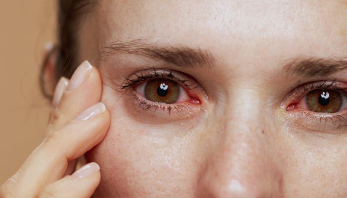
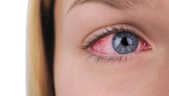
One of the most commonly seen problems in an eye doctor’s office is the red eye. Conjunctivitis, a generic term which describes inflammation of the conjunctiva, the clear membrane which covers the eye, is only one of many causes of red eyes. Many other diseases, from benign to vision-threatening, must be considered and will be described in the following discussion.

Like the tissues which line the inside of the nose and mouth, the conjunctivae are mucous membranes. Conjunctiva covers nearly the entire exposed surface of the eye, as well as the inside of the eyelids. This tissue serves a number of functions, including protection of the eye from injury and infection, as well as production of tear components to keep the eyes moist. Inflammation of the conjunctivae, or conjunctivitis, is a very common problem with many possible causes, including infection, allergy, trauma, and immune reactions. Symptoms vary depending upon the type of conjunctivitis present.

Viral conjunctivitis is the most common form. Often called “pink eye,” this disease is caused by a number of different viruses, often the same pathogens which are responsible for respiratory infections such as the common cold. These viruses are often very contagious, and viral conjunctivitis frequently passes between members of shared environments, such as among children in daycare or between members of a family. Infection can be transmitted through the air by a sneeze, or by contact with contaminated objects such as towels, doorknobs, phones, etc. Ocular changes may be preceded by cold-like symptoms. The eyes often become red, watery, and swolen, with a gritty or foreign body senstation. Crusting of the lashes with clear or yellow discharge is often noted, and the eyelids are commonly stuck shut in the morning. Discharge is usually thin and watery, though may become thicker with mucous. Vision may be blurred slightly. The disease usually peaks after a few days to a week and resolves within 7 to 10 days, much like a cold. No specific therapy is available for viral conjunctivitis, and treatment is supportive. Cool compresses and artificial tear drops provide comfort and cleansing for the eyes. Antibiotics, though often prescribed by physicians, will not treat viral illness and are usually unnecessary. Most important is to remember that the disease may be contagious; thorough and frequent washing of hands will help prevent transmission to others. Rarely, a keratitis, or inflammation of the cornea, will develop, usually with more significant pain and blurred vision. This may require treatment with other medications. An examination by an eye doctor can determine if this is necessary.

Bacterial conjunctivitis is much less common than the viral form. Presentation of symptoms may be similar to viral disease, however pus is typically present. Pus is often white, yellow, or green, and less stringy than mucous. It may reaccumulate quickly if washed or wiped away. The eyes are often very red and inflamed, and may be quite uncomfortable. Unlike viral conjunctivitis, this form is responsive to antibiotics, usually prescribed in the form of eye drops. Recovery is usual within one to two weeks.

Not all conjunctivitis is infectious. Seasonal allergies are commonly associated with ocular symptoms.The primary symptom of allergic conjunctivitis is itching, often severe. The eyes typically appear pink and boggy, tear significantly, and may have a discharge of mucous. Other symptoms of allergy, including runny nose and irritated throat, are often present. Oral antihistamines and other systemic allergy medications are often helpful, however greater relief is often achieved with eye drops specially formulated for ocular allergy. Prescription medications are usually superior to over-the-counter products.

Toxic or chemcal conjunctivitis is due to exposure to certain chemicals or irritants. Any chemcial exposure to the eye should be considered an emergency, requiring immediate and throrough irrigation of the offending substance. Assuming no significant chemical burn is present, care is generally supportive. Occasionally, specific medications are necessary.

Conjunctivitis is often associated with blepharitis and dry eye syndrome. These conditions are covered in separate sections of the library, and will not be detailed further here.

Red eyes, as discussed earlier, do not always signify conjunctivitis. Numerous other ocular diseases and conditions cause the eyes to take on this hue. Inflammation of a deeper layer of tissue, the episclera, will often cause a focal area of redness, at times associated with mild discomfort. This episceritis is generally an isolated condition. The condition resolves spontaneously within a few days to a week with treatment usually unnecessary, although mild steroids can speed recovery.

Inflammation of the sclera, the thick white wall of the eye, can also cause localized or diffuse redness and swelling. Scleritis is often very uncomfortable, described as a deep, piercing pain that may wake an individual from sleep. The eye may be very tender to touch. About half of the time scleritis is associated with autoimmune diseases such as rheumatoid arthritis or lupus. This disease can be serious, potentially leading to loss of vision. Treatment involves steroid eye drops, systemic non-steroidal or steroid medications, and rarely other immunosuppressive drugs.

Redness concentrated in a ring around the cornea is often seen with iritis or uveitis. This inflammation of uveal tissue, which includes the iris, the ciliary body, and the choroid, is not uncommon and has many possible causes. Other frequently occurring symptoms include blurred vision, pain, and sensitivity to light. One or both eyes may be affected, and recurrences are common. Treatment is with steroid eye drops, and occasionally systemic medications if severe.

Keratitis, or inflammation of the cornea, will also lead to redness along the corneal margin. There are many causes of keratitis, both infectious and non-infectious, and diagnosis requires careful examination and, at times, special tests. Treatment depends upon the underlying cause.

Acute angle-closure glaucoma will cause red-eye with blurred vision, halos around lights, and pain. This condition is discussed in the section on glaucoma.

A very common cause of red eye is subconjunctival hemorrhage. In this condition, a small blood vessel breaks, leading to bleeding within or under the conjunctiva. A bright red, bloody appearing spot, sometimes quite large, is noted on the surface of the eye. A mild ache or irritation may be present. These hemorrhages may occur after coughing, sneezing, vomiting, or straining at stool.
They may be more common in individuals on blood thinning medications. Often, no specific cause can be found. This condition can appear quite frightening, however is nearly always benign, the equavalent of a bruise on the skin. The eye will not be injured and vision will not be affected. The blood will clear over a few days to a few weeks, depending upon the extent of bleeding.

Clearly, there are numerous causes of red eyes, some totally benign and some more serious. A thorough eye examination will allow your doctor make the diagnosis and prescribe appropriate therapy. Contact us to schedule an appointment today.

Cataracts and Cataract Surgery

Cataracts

Many believe that a cataract is a growth or membrane which develops over the eye. Actually, cataract describes a cloudiness or opacification of the natural lens of the eye. The lens is found within the eye, just behind the colored structure known as the iris.

Ocular anatomy

Much like the lens of a camera, the eye’s lens functions to focus light entering the pupil upon the retina, a thin membrane in the back of the eye. The retina functions like film, sending what the eye sees on to the brain through the optic nerve. Just as a dirty camera lens will take a blurry photograph, a cataractous human lens can cause a number of visual disurbances, including blurring, haziness, glare, difficulty judging colors, and eventually significant loss of vision in the eye.

For most people, cataracts are simply a matter of age.

Nearly everyone develops cataracts over time, though the rate at which the lens undergoes these changes is quite variable. Some may note significant visual disturbances in their 40s, while others may live into their 80s and never be very bothered by their vision. The reasons for these differences are not well understood. Genetics may play a part, with some simply more succeptible to cataract at an earlier age. Environmental factors, such as exposure to sunlight or certain diets, may one day be found to play a role, however current data regarding the effects of these elements are not conclusive.

While most cataracts are age-related, some are not. Some people are born with cataracts. Others may develop cataracts due to illness or disease, trauma, or use of certain medications. For example, those on chronic corticosteroids such as prednisone often develop lens opacities. By understanding your full medical history, your doctor can determine whether your cataracts are related to any of these factors.

Presently, the only available treatment for cataracts is surgery.

Many often ask if cataracts can be prevented. Given our current lack of understanding of their causes, the simple answer is no. Some eye doctors advocate the use of ultraviolet light-blocking sunglasses. While not definitive, some studies have suggested that ultraviolet light may be partly responsible for cataract progression, as well as for other eye diseases such as age related macular degeneration. The use of sunglasses may therefore help, and certainly will not cause any harm. Similar advice might apply to antioxidants such as vitamins A, C, and E. While studies have produced conflicting information, some indicate that these vitamins may slow cataract growth. Speak with your medical doctor prior to starting any high-dose vitamin program.

Cataract surgery may be right for you if you experience any of the symptoms noted above, such as significant blurring, haziness, or glare. If any of your work or leisure activities are affected by poor vision, a complete eye examination will help your physician determine the cause. Patients with cataracts often complain of difficulty reading or performing close-up work, trouble driving- especially at night due to glare and starbursting around lights, or difficulty following a golf ball.

Modern cataract surgery is performed without discomfort on an outpatient basis. Gone are the days of large incisions, multiple sutures, and prolonged bedrest. Today, incisions are tiny, typically measuring only three millimeters, or about an eighth of an inch. Stiches are seldom necessary, and recovery, both physical and visual, is rapid. Many patients drive and return to their usual level of activity the day following surgery.

The surgical procedure: phacoemulsification

The details of cataract surgery, known as phacoemulsification, will be described next. Please visit our Outpatient Surgery Center page for additional information about your surgical day experience in our state-of-the-art facility.

Once adequate anesthesia has been achieved, the area around the operative eye will be cleansed for sterility and a surgical drape will be placed. A small device known as a speculum will then be placed to keep your eye open during the procedure. There will be no discomfort. A small incision is then made in the cornea, or clear window at the front of the eye, and a viscous gel is injected into the eye in order to maintain space and protect the other ocular structures during surgery.The “main” corneal incision is then carefully made using an extremely sharp and precise blade made of diamond. Next, a circular opening is created in the front of the thin membrane, or capsule, which surrounds the lens, and fluid is injected inside to separate the entire lens from its attachment to the capsule. The cataractous lens is then removed using phacoemulsification, the amazing technology behind modern cataract surgery. The phacoemulsification machine produces ultrasound waves within a tiny handpiece, pictured to the left, which is inserted into the eye. This causes the metal tip of the instrument to vibrate very rapidly, up to 40,000 times each second, dissolving the cataract material which is then aspirated out of the eye. Additional instruments are used to clean all the cataract from the lens capsule, which is then refilled with more viscous gel. An artificial, permanent lens implant, made of a type of plastic, is then inserted into the capsular “bag,” taking the place of your natural lens. This implant, which helps focus the incoming light on your retina, is held in place by two haptics, or curved arms. Once the implant is in place, the incisions are closed, usually without stitches, and the surgery is complete.

Pre-operative measurements and intraocular lens implants

Prior to surgery, your eye will be carefully measured for the proper strength of lens implant. The implant can be chosen to focus your eye at any distance you desire. While most choose to focus at distance, some prefer to see most clearly at near, particularly if they have always been myopic, or nearsighted. At Fishkind, Bakewell Maltzman & Hunter we perform these measurements with the Zeiss IOL-Master, one of the most advanced, accurate devices for this purpose, which uses a laser to scan the eye in just seconds. While these calculations are extremely accurate in most cases, the expected results are not always achieved. Most patients obtain vision good enough to drive without glasses. Rarely, the post-operative refraction (eyeglass prescription) will be significantly different than expected. In these unusual cases, additional surgery, including removal and replacement of the lens implant, may be required to achieve the desired result.

The majority of lens implants inserted during cataract surgery are considered ‘monofocal,’ meaning that they focus light at only one location, usually in the distance. This means that reading glasses will be required for close-up tasks such as reading- without them near vision will be blurred. Other options exist, however, including lenses that correct astigmatism (see below) and that provide clear vision from distance to near. You can discuss these options with one of our physicians during your evaluation. Please click here to read in greater detail about lens implant options available.

Management of astigmatism

Astigmatism is a form of refractive error in which perpendicular lines are focused at different distances within the eye. Significant astigmatism occurs in as many as one in three people and can cause blurred vision, visual fatigue, and squinting if not corrected with glasses or contact lenses. In early life, most astigmatism is due to an irregular shape of the eye, in which the cornea is steeper in one meridian than the other. As we age, many develop astigmatism due to irregular thickening of the lens as cataracts develop. This latter form of astigmatism will disappear when a cataract is removed, however any remaining corneal astigmatism will blur vision, increasing the likelihood that glasses will be necessary for clear vision postoperatively.

Fortunately, for those who desire the best possible uncorrected distance vision after surgery, astigmatism can be reduced or eliminated surgically at the time of the cataract operation with a special lens implant known as a “toric IOL.” Like eyeglasses, which are crafted with cylindrical lenses placed in a specific axis to correct one’s particular degree of astigmatism, a toric implant with the correct cylindrical power is carefully rotated into the precise position within the eye to neutralize the corneal astigmatism.

Posterior capsular opacification

Once removed, a cataract does not return. However, the back side of the lens capsule, which remains intact at the time of surgery in order to support the implant, can become cloudy over time. This opacification, sometimes referred to as a “secondary cataract,” can affect vision much the way the cataract once did. If this occurs, a brief, painless laser procedure performed in the office can clear the opacity from the implant. The capsular opacification does not recur once this is performed.

Modern cataract surgery is safe, effective, and capable of rapidly improving your vision. Don’t let poor vision slow you down. Call or contact us today to schedule an appointment with one of our doctors to find out if cataract surgery can help you.

Blepharitis

What is blepharitis?

Blepharitis, which literally means “inflammation of the eyelids,” is an extremely common condition in which the eyelids become red and swollen, often leading to symptoms of burning, itching, irritation, foreign body sensation, sensitivity to light, and blurred vision. Although the condition is not an actual infection, toxins released by bacteria which normally live within the skin of the eyelids contribute to the associated inflammation. Blepharitis is often divided into two forms- anterior and posterior.

In anterior blepharitis, inflammation is most pronounced toward the front of the eyelids, involving the glands associated with the eyelashes. Usually caused by an overgrowth of staphylococcus bacteria, this condition is often seen in children and young adults. Bacterial toxins irritate the surface of the eye, causing conjunctivitis, or inflammation of the membrane which lines the eyeball. In more severe cases, the cornea becomes involved in a condition known as marginal keratitis. White inflammatory deposits develop in the periphery of the normally clear cornea, leading to significant pain, foreign body sensation, blurred vision, and often extreme sensitivity to light.

In posterior blepharitis, more commonly found in adults, inflammation involves the oil-producing Meibomian glands which line the back of the eyelid margins. The normal, smoothe oils produced by these glands, which serve to lubricate the eye, become thick and greasy, clogging the gland openings. The thick secretions are very irritating to the ocular surface, causing all of the symptoms noted above. This condition is often associated with the skin disease rosacea, in which the oil glands of the face become inflamed, leading to red, inflamed skin over the brows, nose, and cheeks.

How is blepharitis treated?

Blepharitis tends to be a chronic condition, waxing and waning with intermittent flare-ups. While not curable, most symptoms can be adequately controlled with proper care. Treatment depends on the type and degree of blepharitis present.

The mainstay of treatment for all forms of the disease is the use of hot compresses and cleansing of the eyelids regularly. This is accomplished by placing a moist, very warm washcloth or other form of hot pack over the closed eyes for a few minutes in order to loosen oils, dead skin, and excess bacteria, followed by gently massaging the upper and lower lids with either the cloth or a commercial eye scrub product to remove the remaining debris. This is best performed at least twice daily- upon waking and before bed.

Additionally, your doctor may prescribe medications. In some cases, antibiotic drops or ointments may be used to help limit the growth of eyelid bacteria. Steroid drops or ointments, alone or in combination with an antibiotic, may also be used to control inflammation and reduce redness and swelling. These medications are typically prescribed for a short, specific period of time, as side effects limit their extended use.

For more long-term control, other medications may be used. Doxycycline or minocycline, medications which have both antibiotic and anti-inflammatory properties, may be taken daily as a pill for an extended period of time in order to control the inflammation associated with the disease. Because these medications are taken orally, systemic side effects such as upset stomach and diarrhea can be a problem. In such cases a topical (eyedrop) antibiotic called Azasite® (azithromycin) can be used with similar success. Restasis® (topical cyclosporine) eye drops, a commonly used treatment for dry eye problems, have also been found helpful in controlling symptoms.

The symptoms of blepharitis are common in many other eye disorders, some benign and some more serious. A thorough eye examination will allow your doctor to distinguish among these conditions and provide the correct diagnosis. If your eyes burn, itch, ache, or get red frequently, call us today to schedule and appointment.

Now Offering Eyhance and Vivity Premium Lenses

If your vision is currently impacted by cataracts, it is essential to schedule an appointment with one of our ophthalmologists for proper treatment. We offer cutting-edge technology to help patients improve or manage their vision problems. Cataracts can be removed with surgery and a new Intraocular Lens (IOL) is implanted to replace your natural lens.

The Eyhance and Vivity premium lenses are two of the state-of-the-art IOLs that provide patients with significantly improved vision. That is why our ophthalmologists at Oracle Eye Physicians & Surgeons in Tucson, AZ, offer Eyhance and Vivity premium lenses for individuals interested in their best corrected vision after cataract surgery.

Why Choose Eyhance and Vivity Premium Lenses?

The traditional IOLs used to replace natural eye lenses are generally able to provide either near or distance vision and then glasses may be needed after surgery for additional correction. The new premium lenses allow for more clarity due to recent advancements in eye care technology. These lenses have recently been approved for use in the United States. Vivity is the first non-diffractive Extended Depth of Focus (EDOF) lenses, and Eyhance is one of the first enhanced monofocal lenses in the US. These premium lenses will also allow you to see more clearly and with more light. Enhanced monofocal lenses will extend your depth of focus and expand your range of vision.

Contact Us for Cataract Eye Surgery from Our Tucson Ophthalmologists

You should consider Eyhance and Vivity Premium eye lenses if you are interested in broadening your scope of vision and seeing more clearly. If you are undergoing cataract eye surgery soon and live in the Tucson, AZ, area, contact the professionals at Oracle Eye Physicians & Surgeons to discuss premium lenses for better vision. Our ophthalmologists are committed to providing quality eye and vision care to patients at our Tucson locations. Contact us through our website or call us at 520-293-6740 to learn more about how we can help.

Light Adjustable Lens for Cataract Treatment

If you are suffering from cataracts and wish to have cataract surgery, you should consider a light adjustable lens (LAL) as one of your options. This lens allows your vision to be customized after cataract surgery. At Oracle Eye Physicians & Surgeons in Tucson, AZ, our experts can determine if the light adjustable lens is right for you.

How Does a Light Adjustable Lens Work?

Cataracts occur when the natural lens in your eye becomes cloudy. This blurs and darkens your vision, making it hard for you to see. This is one of the leading causes of impairment in vision, and it is common in people as they age. The only real treatment for cataracts is to remove this cloudy lens and replace it with an artificial one, known as an intraocular lens, or IOL. Cataract surgery can correct many other visual problems as well, such as farsightedness, nearsightedness, and astigmatism.

Before cataract surgery, our ophthalmologist will use detailed measurements of your eye to select the type of intraocular lens that will give you the best possible vision after surgery. However, no matter how many measurements are taken, it is still only a best estimate and not a perfect science. Every eye heals a little bit differently after surgery, so some patients do not experience perfect vision. If the visual target is not reached, this can typically be corrected with contacts, glasses, or even additional surgery.

With a light adjustable lens, we are able to adjust your vision after cataract surgery. This allows you to have a lens that has been customized specifically for your eyes. The lens is made of a special photosensitive material that changes in response to UV light. This allows the lens to change its curvature and refractive power. Once your surgeon completes the final lens adjustment, the entire lens is exposed to light that locks it in.

The light adjustable lens is a great option for patients who have had previous refractive surgery or wish to customize vision after cataract eye surgery. Our eye surgeon will work with you to determine if you are a candidate for the LAL option and discuss surgery with you. After surgery, the surgeon completes detailed measurements and adjustments with you to customize the LAL for your best vision outcome.

Visit Our Ophthalmologists in Tucson

If you are suffering from cataracts, we can offer you a long-lasting solution. At Oracle Eye Physicians & Surgeons in Tucson, AZ, our eye surgeons offer light adjustable lenses to give you a customized result after eye surgery. We can answer any questions you have about the light adjustable lens and cataract surgery. To learn more about our services or to schedule an appointment, call us today at 520-293-6740 or send us a message online.