Pseudoexpoliation Glaucoma
As noted in the general glaucoma review, the vast majority of people with the disease have the open angle type, and most of these have what is known as primary open angle glaucoma, or POAG. The main feature of POAG, aside from elevated intraocular pressure (IOP) and visible damage to the optic nerve, is an otherwise normal eye. In other words, there are no other identifiable abnormalities to explain either the pressure or optic nerve damage. The cause or causes of POAG are largely unknown, though numerous genetic defects are being studied for their role in the disease.
A significant number of patients with open angle glaucoma have special forms of the disease, known as secondary open angle glaucoma. In these conditions, there are identifiable abnormalities responsible for the elevated intraocular pressure and associated damage. One such condition, pseudoexfoliation glaucoma, is described below.
Pseudoexfoliation Syndrome
Also known as Pseudoexfoliation Glaucoma, pseudoexfoliation glaucoma (PXFG) is probably the most common type of secondary open angle glaucoma, accounting for 10 to 25 percent of glaucoma worldwide. The disease is caused by production of an abnormal material within some of the tissues of the eye. This material ultimately ends up within the eye’s drainage system where it causes damage, essentially clogging the channels and raising intraocular pressure. While the exact cause of PXFG is not known, strong evidence suggests that one or multiple genetic abnormalities are responsible. Indeed, a recently identified gene has been found to be significantly associated with pseudoexfoliation. Further research will help us learn the exact role of this gene in the disease.
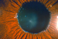
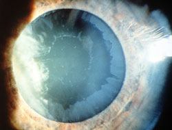
Diagnosis of PXFG is made by identifying the abnormal material, which looks a bit like dandruff flakes, on the iris or lens. It is important to have the pupils dilated, as the material may be missed otherwise. Visualizing the material can be difficult, particularly early on in the disease, and it may not be found until years after the initial diagnosis of glaucoma is made. The presence of pseudoexfoliation, and the associated risk of glaucoma, increases with age.
Although very similar to POAG, PXFG does have a few unique features:
- While elevated IOP and optic nerve damage tends to occur in both eyes symmetrically in POAG, it is often markedly asymmetric in PXFG, with one eye affected much more than the other. Indeed, some patients with PXFG appear to have glaucoma in only one eye.
- IOP fluctuates more widely and pressures run higher in PXFG. Furthermore, IOP is often more difficult to control with medications than in POAG. Medical treatment will often require more than one drug to control IOP.
PXFG is treated similarly to other types of open angle glaucoma. Medications are usually the first option. However, as noted above, medications are often less effective than in POAG. Laser trabeculoplasty can have a very good effect in PXFG, often dropping the IOP significantly. The drop in pressure from trabeculoplasty is temporary, however, and IOP may begin to elevate again within a few years. Ultimately, surgical intervention may be required in at least one eye.
Not every patient with pseudoexfoliation will develop glaucoma. There is about a 70 to 85% chance that elevated IOP and glaucoma will occur, and the risk increases with age. If pseudoexfoliation material is identified in your eye during an examination, observation 2-3 times per year is indicated to watch for signs of rising pressure.


