Mon - Fri: 8:00 am - 5:00 pm, Sat & Sun: Closed
Give Us A Call Today!
(520) 355-7501
Give Us A Call Today!
(520) 293-6740
Lasik Eye Surgery in Tucson Lasik Eye Surgery in Tucson

“EXCELLENT SURGEONS THAT ARE CARING PEOPLE, TOO!”

Please Fill Out the Form Below or Call Us to Make an Appointment

The #1 Glaucoma Management in Tucson and Nearby Areas

Glaucoma is a group of eye disorders that can lead to progressive loss of vision, often without any symptoms. Visual loss results from damage to the optic nerve, which transmits signals from the eye to the brain, much like an electrical cable. Once nerve fibers are damaged by glaucoma, the information they supply can no longer reach the brain. Peripheral vision is usually lost first.

Because central vision is preserved until late in the disease and pain is usually absent, most people with glaucoma don’t realize that anything is wrong. 

To ensure that you have the greatest chance of success, trust a qualified team to care for your eye health.

If you’re in the Tucson and Oro Valley area, you will find an experienced eye surgeon at Fishkind, Bakewell, Maltzman & Hunter Eye Care and Surgery Center.

5-Star Rated Glaucoma Management in Tucson.

Hear What Our Customers Say

A general overview

Diagnosing For Glaucoma Management in Tucson

Unfortunately, the diagnosis is glaucoma is not always straightforward, even after all testing is performed. There is significant variation in the appearance of normal optic nerves, with many healthy nerves looking a bit suspicious for glaucoma. Because glaucoma is usually a very slowly progressive disease, it is not uncommon for patients to be watched carefully without treatment while the diagnosis of glaucoma is considered. These ‘glaucoma suspects’ may be followed for many years before a final diagnosis is made and treatment is initiated. If suspicion is relatively low, ‘watchful waiting’ avoids the potential risks and side effects of treatment.

Step One: Medical History and Eye Examination

Your physician will perform a complete medical history and eye examination in order to determine your risk factors and search for signs of disease. The entire eye will be carefully evaluated, with special attention paid to the optic nerve, the structure damaged by glaucoma. The optic nerve is viewed through your dilated pupil using a special lens and an ophthalmic microscope. Gonioscopy may also be performed, in which a special mirrored lens in placed on your eye in order to directly examine the drainage angle and trabecular meshwork. The thickness of your cornea may also be measured, as an abnormally thick or thin cornea can affect the accuracy of intraocular pressure measurement. After your exam, if glaucoma is suspected, additional tests may be recommended.

Step Two: Stereoscopic Optic Nerve Photographs

Stereoscopic optic nerve photographs are taken with a specialized camera system, providing true, three-dimensional images of the optic nerves. These photos can be repeated over time and examined for signs of progressive nerve damage. In order to obtain clear photos, pupillary dilation is required. This examination usually takes about 45 minutes, including the time required for dilation.

Step Three: Visual Field Testing

Visual field testing is conducted to evaluate the function of the optic nerve. As glaucoma damage progresses vision is lost, typically beginning in the periphery and moving toward the center. Computerized visual field devices flash a series of lights in your peripheral vision in order to map out any areas of visual loss. Again, by repeating this test over a period of time, stability or progression of the disease can be ascertained and treatment adjusted as necessary. Completion of this test can take anywhere between 20 and 45 minutes, depending upon the exact type of examination your doctor orders. Your eyes will not be dilated for a visual field.

Step Four: Ocular Coherence Tomography

Ocular Coherence Tomography, or OCT, uses a laser to create a three-dimensional image of the optic nerve and surrounding retina, which is then analyzed by a computer. This allows your physician to better evaluate the structure of the fibers of the optic nerve, which are altered by glaucoma. Changes in the structure of the optic nerve occur early in glaucoma, often preceding any visual changes, and identification of these changes may be the best way to diagnose the disease. By repeating this exam over time, the success of glaucoma treatment can be monitored. The performance of this test usually does not require dilation and takes about 15 to 20 minutes.

Step Five: Treatment
Treatment for glaucoma is individualized for each patient’s specific condition. When required, three basic options are available, all of which serve to lower the intraocular pressure. Initial therapy has traditionally been medications, applied topically as eye drops. Medications lower pressure by either decreasing the production of fluid within the eye or by opening the drainage system to allow more fluid out. There are numerous classes of medications available, each with its own set of side effects. Your doctor will suggest medications for you based upon their safety and efficacy, taking your general health and use of other medications into account.When medications or laser procedures fail to adequately control intraocular pressure (IOP), or if side effects prevent the continued use of medications, surgery becomes necessary to achieve the desired level of IOP. Surgery for glaucoma lowers IOP by either increasing the flow of fluid out of the eye or by decreasing the production of intraocular fluid.
Glaucoma of the eye
Glaucoma Risk Factor
Risk of Glaucoma in Tucson

What Are the Risk Factors of Glaucoma?

A number of risk factors for the development of glaucoma have been identified:

  • High intraocular pressure: IOP above 21 is considered high, and with increasing pressure comes an increased risk of glaucoma. However, not all high pressure will lead to glaucoma. 

  • Race: African Americans are five to six times more likely than Caucasians to be affected by the disease, which tends to be more severe and progress more quickly in this population. Latinos are also at greater risk than Caucasians. The reasons for these racial differences are not well understood but are most likely genetic.

  • Family History: History of glaucoma, particularly in a first-degree relative, i.e. parent or sibling, increases one’s risk. The genetics of glaucoma, however, are usually not simple, and there is no way to predict which family members will be affected. 

  • Age: Glaucoma is uncommon under 40, but risk increases with each decade of life.

  • Blood Pressure: High blood pressure has been associated with increased intraocular pressure in some studies, but it remains unclear exactly how significant this association is clinically.

    Low blood pressure may be a risk factor for glaucoma progression, particularly in patients with low tension glaucoma, a form of open-angle glaucoma in which the pressure is in the normal range, often quite low.

  • Sleep-associated breathing disorders: Disorders such as sleep apnea, may be a risk factor, particularly for low tension glaucoma, again due to abnormal blood flow to the optic nerves.

Other possible risk factors include diabetes and myopia (nearsightedness), however studies have provided inconsistent results as to the degree of association with these factors.

What Types of Treatments are Available in Glaucoma Management?

Treatment for glaucoma is individualized for each patient’s specific condition. There are three basic options available, all of which serve to lower the intraocular pressure. After medication, Laser Surgery and Micro-Invasive Glaucoma Surgery, or MIGS procedure, are options to treat your glaucoma.

  • Medications

    Medications lower pressure by either decreasing the production of fluid within the eye or by opening the drainage system to allow more fluid out. There are numerous classes of medications available, each with its own set of side effects. Your doctor will suggest medications for you based upon their safety and efficacy, taking your general health and use of other medications into account.

  • Micro-bypass stents

    This is a Micro-Invasive Glaucoma Surgery, or MIGS procedure, attempting to reestablish good flow through the eye’s normal drainage system. Currently, there are two types of stents available for implantation in the United States, the Glaukos® iStent®, which is now in its third generation is known as the iStent Inject®, and the Ivantis® Hydrus® microstent.

  • Ab-Interno Canaloplasty (AbIC)

    This MIGS procedure dilates and cleans out Schlemm’s canal. A tiny catheter, or hollow tube, is inserted into the canal through the trabecular meshwork and passed 360 degrees around the entire eye, breaking adhesions and scar tissue that have formed over time.

  • Goniotomy

    This MIGS procedure involves either cutting into or removing a variably sized section of the trabecular meshwork, allowing aqueous humor a more direct pathway into Schlemm’s canal. Overall, efficacy is similar to that of AbIC, though depends upon the extent of meshwork treated.

  • Trans-scleral Stents

    This procedure bridges the gap between MIGS procedures and traditional filtering surgery. Like filtering surgeries, aqueous humor is diverted from inside to outside the eye via a bypass through the sclera, forming what is known as a “filtering bleb.” However, much less cutting and manipulation of tissues is required when implanting these devices, with considerably shorter operative and recovery times.

  • Laser Trabeculoplasty

    This laser treatment relieves intraocular pressure by stimulating the internal drainage system allowing adequate outflow of fluid from the eye. There are two types of laser trabeculoplasty procedures: argon laser trabeculoplasty (ALT) and selective laser trabeculoplasty (SLT)

  • Laser Peripheral Iridotomy (LPI)

    This laser treatment makes a small opening in the peripheral iris (the colored part of the eye). This changes the fluid dynamics in the eye and “opens” the angle. If a patient already has acute angle closure glaucoma, the small opening that is made in the peripheral iris allows fluid to drain more normally and also lowers the eye pressure.

    See Now – Pay Later! Finance Your LASIK Procedure with CareCredit!

    As a service to our patients, we are pleased to offer the CareCredit card, the nation’s leading patient payment plan. With CareCredit you can finance 100% of your procedure and there are no upfront costs, no annual fees, and no pre-payment penalties. So, you can begin your refractive or elective procedure today and conveniently pay with low, monthly payments.
    Tucson LASIK Eye Surgery Center

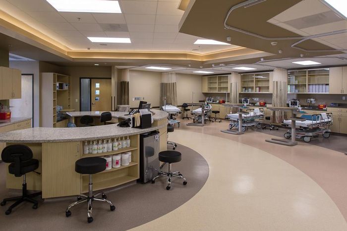
    Why Our Facility Is a Great Glaucoma Management Center for You

    We know that you have a choice of teams and locations for your care. Our team of experts look forward to providing the highest quality care. Our experienced team, high-tech equipment, and personalized care provide you with the best outcomes for your vision. 

    FAQ

    Glaucoma Questions & Answers

    What causes Glaucoma?

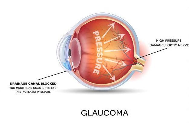
    Scientist are not entirely sure what exactly causes Glaucoma. Ultimately the passages that allow the fluid within your eye to drain are blocked. Because of that, fluid builds up in your eye and causes an increase in pressure on the optic nerve. The optic nerve and blood vessels are damaged by the increased pressure, which can result in vision loss.

    Will Glaucoma Make Me Go Blind?

    Glaucoma can cause blindness, but it can be prevented. If it is diagnosed at an early stage, the damage can be controlled. If treatment is delayed, you are likely to lose peripheral vision first and then the central vision, which may result in complete blindness.

    How can I tell if I have Glaucoma?

    Glaucoma often does not have obvious symptoms. Additionally, symptoms may not show until it is already too late. 

    If you have open-angle Glaucoma, the initial symptom might be vision loss – which is a sign of damaged optic nerve.

    If you have closed-angle closure Glaucoma, initial symptom may be blurry vision. You might also see rings or halos around lights. You may experience pain or strain in your eye and they may also get red.

    What are the Types of Glaucoma?

    Glaucoma is usually broken down into two major categories: open-angle glaucoma and closed-angle glaucoma. 

    In open-angle glaucoma, the drainage canals are correctly formed but are slow clogging, resulting in increased eye pressure.

    In closed-angle glaucoma, the entrance to the trabecular meshwork becomes blocked by the iris, which assumes an abnormally forward position. This causes the drainage canal to become narrow and eventually close. Unlike symptom-free open-angle glaucoma, this condition, sometimes called “narrow-angle glaucoma,” often presents suddenly, with pain, red eye, tearing, and colored haloes around lights. Vision can be lost quickly, and urgent treatment is required. 

    Are there Restrictions After Surgery?
    The degree of allowable physical activity, including the ability to drive in the days following surgery, depends on each patient’s unique situation and must be judged individually. In general, strenuous activity, heavy lifting, and bending over should be avoided for the first one to two weeks.
    Is Surgery a Permanent Solution?
    Drainage device surgery is generally an effective procedure for IOP reduction, with a 60 to 80% chance of maintaining intraocular pressure at the desired level after one year. Unlike trabeculectomy, in which roughly half of patients need no additional IOP-lowering medication, drainage devices are more likely to require supplemental glaucoma medication to maintain adequate IOP levels. As the years pass it is not unusual for IOP to begin to increase, possibly necessitating the use of additional medications, or additional surgery. In approximately 20 to 40% of cases, surgery fails to adequately control the IOP beyond the first year or two. Additional surgery may then be performed to reach the desired level of pressure within the eye. A second drainage device might be implanted, or other options can be considered. pressure and prevent the further loss of vision. In some cases, glaucoma medications may be eliminated, however many patients will need to continue all pre-treatment eye drops to maintain adequately low IOP. Trabeculoplasty is not 100% effective at lowering intraocular pressure. Results often vary depending upon the type of glaucoma, number and type of eye drops being taken, and other conditions affecting the eye. Your surgeon will discuss your specific situation and will provide appropriate guidance. Patients who have undergone drainage device surgery have an increased lifetime risk of developing an infection inside the eye. Known as endophthalmitis, this kind of infection can seriously harm the eye and lead to loss of vision. The increased risk is due to the opening made in the eye; just as fluid can more easily escape the eye, bacteria can more easily enter. While this risk is generally low, anyone who has undergone drainage device surgery is advised to contact their ophthalmologist immediately if the operated eye ever becomes significantly red or painful, or if vision suddenly decreases. For this reason, the use of contact lenses after drainage device procedures is discouraged.
    Is Surgery Safe?
    All surgical procedures carry some risk of complications. Some risks are common to all procedures and patients, and others are more specific to certain types of surgeries or to patients with particular conditions. A thorough explanation of complications will be provided with a surgical consent, should you choose to have surgery, and your physician will review the specific issues you may face based upon your unique circumstances. Complications from SLT are uncommon, with mild corneal or intraocular inflammation occurring infrequently, potentially requiring additional treatment. Complications causing loss of vision are exceedingly rare.
    Contact Us Today to Schedule Your Appointment